

Table of Contents
Introduction
Even though summer is coming close, you can still pack up your most fashionable bright summer acrylic nail designs later. In the upcoming weeks, you can make your consumers seem more knowledgeable and stylish by providing fresh and adorable acrylic designs. Combining acrylic nails, powders, finishes, and pin-above art styles will give each customer who comes to your salon a pair of extraordinary pensionaries.
We have compiled a list of some adorable summer acrylic nail ideas that your customers will enjoy and find suitable for any event, occasion, or style to assist them in making the most of the remaining beach days and holidays. It will help them make the most of the remaining opportunities to go to the beach.
What is a Bright Summer Acrylic Nails?
A powdered polymer and a liquid monomer component are mixed to create a molecular paste when making acrylic nails. You can paint this paste onto a client’s natural nail, and the acrylic nail is fused to harden it. Afterward, you can hang them out somewhere and let them dry naturally without subjecting them to heat further.
These nails provide your clients the length and shape they’ve always wanted and offer you the ideal canvas onto which you may paint some delightful decorations. Even though they need upkeep and care, your clients will be willing to go to great lengths to guarantee that they maintain your outstanding work for as long as possible since these nails will look amazing.
Your clients can enjoy their new manicures up to the end of summer. Since acrylic nail designs often last anywhere from six to eight weeks. You or your customer may feel overwhelmed by the fantastic alternatives available. Still, there is no need to worry about it since we have you covered with a rundown and recommendations for some of the season’s most popular silhouettes and designs.
How Long Do Acrylic Nails Last?
Cosmopolitan says a complete set of artificial acrylic nails may last up to eight weeks if properly cared for. When you fill in the gaps that will appear due to the extension of your natural nails, you must see your go-to manicurist every two to three weeks. According to pros, you shouldn’t wait more than three weeks between getting your fillings and treating your nails.
The increased pressure on your natural nails generated by the larger surface area will be relieved when you do this. In addition to that, it helps ward off fungal infections.
First, Choose the Shape
Before you start your acrylic summer trip together, you should ensure that your client is well-versed in the several types of nail forms accessible to them.
Almond Bright Summer Acrylic Nails
The nail wall of almond nails is filed down to a point, creating a rounded almond shape at the tip of the nail. Because of their inherent weakness, natural nails often struggle to keep almond-shaped nail shapes on their own. As a result, acrylics are an excellent choice for customers who are adamant about maintaining this shape on their nails.
Coffin Bright Summer Acrylic Nails
The coffin acrylic is not quite as horrible as its name might be the main one to believe it is. Longer acrylic nails, which have been gaining popularity recently, may create the appearance of longer fingers and a smaller profile for your consumer.
The coffin-shaped nail is thin at the base, and it expands at the tip, giving the appearance of wider fingers than the wearer has (which ends up making it look like its namesake or, perhaps more appealingly, like a ballerina slipper).
If you want the luxurious look preserved for as long as possible, inform your customers that they need to handle this particular acrylic with more care since it is more brittle than the others.
Mountain Peak
Your consumers will appreciate that these acrylics give elevation and attitude, enabling them to carry the warm temps and positive sentiments of summer with them wherever they go. Mountain peak nails have a broad base that tapers to a somewhat curved tip and are called for the mountain modeled after.
It should come as no surprise that famous people like Lady Gaga and Rihanna have experimented with this type of manicure, given its many options for creating dramatic and complex nail art.
Here are some Adorable Outfit Ideas for the Bright Summer Acrylic Nails
You and your client are now at the point where you may experiment with some new and cute summer styles. Before settling on one, you and the person you are selling need to investigate each choice that is open to you.
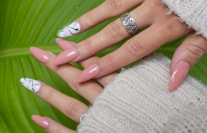
Glossy Summer Floral Nails
Savoring ice cream on a hot summer day, these glossy nude nails adorned with stunning floral prints become your stylish sidekick. Capturing the essence of nature’s beauty and reflecting the sun’s rays, they add a touch of magic to your fingertips, making every day a delightful adventure filled with laughter and sunshine.
Ombre Orange Summertime Nails
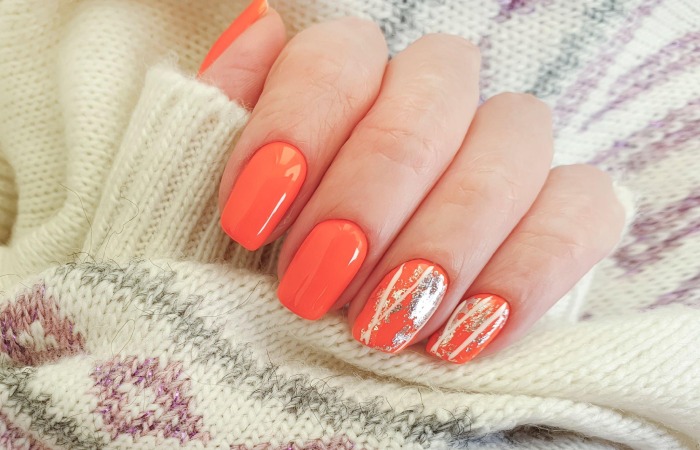
Unleash your inner Egyptian royalty with these gorgeous ombre orange nails, where the essence of summertime intertwines with the allure of old luxury. Long golden flakes adorn the vibrant orange gradient, reminiscent of the treasures hidden within the sands of time. As the sun’s rays glisten upon your fingertips, they unveil a mesmerizing play of light and color, weaving a tale of mystery and majesty. Embrace the season’s warmth while exuding an air of regal splendor that transcends the ages.
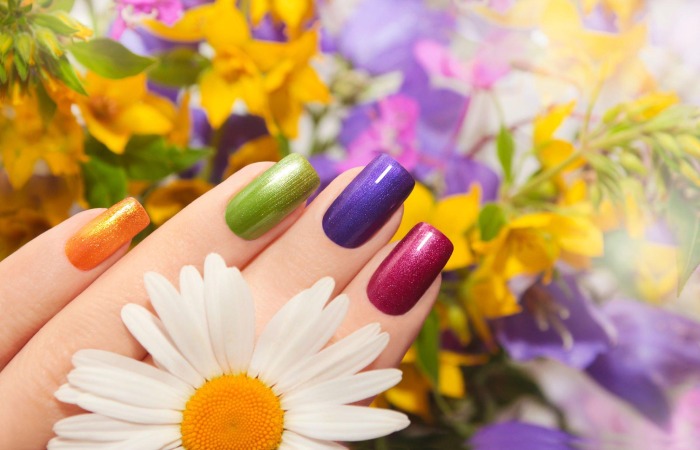
Playful Summer Chrome Nails
These vivacious nails feature an eye-catching chromatic design impeccably for soaking up the sun and making memories that will last a lifetime.
Let your right hand take center stage with stunning shades of green, from the thumb to the ring finger, while a delightful pop of pink graces your pinky. Meanwhile, your left hand flaunts a flirty and fabulous array of pink hues, with a surprising twist of green on the pinky finger. The real showstoppers, however, are your index fingers – each adorned with a checkered pattern that interweaves the corresponding colors, creating a mesmerizing and playful effect.
French Tip
The French tip nail design has been a customer favorite for many years. So there is no need to forsake something performing so well to delight customers repeatedly. Put the final touch on this chic acrylic by adding a few colorful accessories at the ends to create a casual look perfect for summer’s unofficial conclusion.
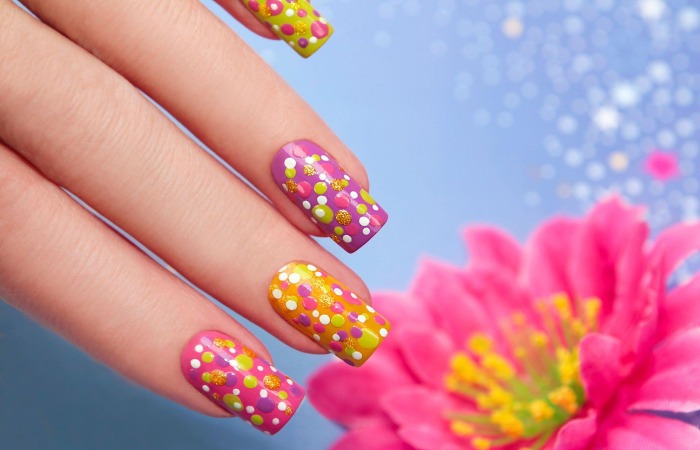
Bright Polka Dot Nail Art
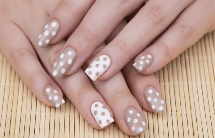
The polka dot nail art’s simplicity and easy application make this a classic and a firm favorite no matter the season. However, choosing bright and saturated colors can give your manicure a distinctly summery feel. There are unlimited options for getting creative, including experimenting with different size dots and the angles of your application.
Bright Floral Nail Art
We love floral nail art because there are many options, letting you experiment with colors, sizes, placements, and even shapes. This is also one of the most symbolic choices because flowers are associated with beauty and growth — so what better way to channel these things than with a super cute and colorful design? Bring attention to your hands and show you’re an individual.
Conclusion
Practice painting colorful bright summer acrylic nails to prepare for the following summer. Our suggestions might be helpful. Choose the second option to look and feel like your fabulous self without sacrificing time or energy. When you fire the trigger, though, you’re the one who has to deal with the consequences. With the information you’ve provided, it’s up to you to decide what to do next. Check to see whether it fits in with the style you’ve been cultivating. We are happy to provide weekly reports that include everything important to you. This article summarizes the most recent developments in nail art for your reading convenience.


